
What is Echocardiography?
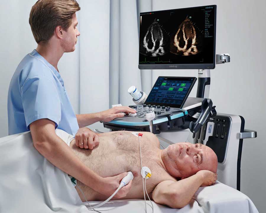
Cardiology practices are at the forefront of treating and managing cardiology conditions and rely heavily on echocardiography due to its portability and non-invasive method of understanding the structure and function of the heart. This includes the diagnosis of various heart conditions like valve diseases, congenital heart defects, heart muscle damage, and can also be used to monitor treatment efficacy.
\r\nCardiology practices continue to be a vital part of the healthcare provider network because they can serve patients’ needs within the community and offer one convenient place to receive cardiac care. Unlike inpatient echocardiography, typically used for patients with more acute and critical heart conditions, office-based echocardiography is used for patients with suspected or known heart conditions requiring routine monitoring or preventive care.
\r\n
Seventy years ago marked an important moment in medical history with the emergence of echocardiography. Echocardiography is a groundbreaking modality that uses ultrasound to visualize the heart. It all began with the publication of the first ultrasound image of the human mitral valve.1 Today, echocardiography has become an indispensable technique in clinical practice, integral to the daily operations of over 2,500 active cardiology physician group practices across the United States.2
Cardiology practices are at the forefront of treating and managing cardiology conditions and rely heavily on echocardiography due to its portability and non-invasive method of understanding the structure and function of the heart. This includes the diagnosis of various heart conditions like valve diseases, congenital heart defects, heart muscle damage, and can also be used to monitor treatment efficacy.
Cardiology practices continue to be a vital part of the healthcare provider network because they can serve patients’ needs within the community and offer one convenient place to receive cardiac care. Unlike inpatient echocardiography, typically used for patients with more acute and critical heart conditions, office-based echocardiography is used for patients with suspected or known heart conditions requiring routine monitoring or preventive care.
Advantages of Echocardiography in Cardiology Practices
- \r\n
- Convenience – access to office-based ultrasound exams in cardiology practices means that patients do not have to travel to multiple hospital visits to receive vital diagnostic care. Practices can move ultrasound machines from exam room to exam room, offering accessibility and shorter wait times. \r\n
- Faster Diagnosis and Treatment – Cardiac Ultrasound provides real-time information about a patient's heart, reducing the time to obtain a diagnosis compared to other imaging modalities. Cardiac Ultrasound not only facilitates patient triage, but it can quickly provide diagnostic confidence to initiate patient care. \r\n
- Continuity of Care - In addition to detecting the presence of cardiac conditions, regular ultrasound scans can be used to monitor patient’s disease progress or recovery. Private office ultrasound also facilitates direct communication between cardiologists and patients, which may result in better compliance with physicians' instructions and better outcomes. \r\n
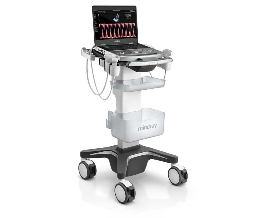
Office-based echocardiography provides a portable, accessible and efficient tool for cardiologists to diagnose and treat heart conditions, bringing significant benefits to practices and patients alike, including:
- Convenience – access to office-based ultrasound exams in cardiology practices means that patients do not have to travel to multiple hospital visits to receive vital diagnostic care. Practices can move ultrasound machines from exam room to exam room, offering accessibility and shorter wait times.
- Faster Diagnosis and Treatment – Cardiac Ultrasound provides real-time information about a patient's heart, reducing the time to obtain a diagnosis compared to other imaging modalities. Cardiac Ultrasound not only facilitates patient triage, but it can quickly provide diagnostic confidence to initiate patient care.
- Continuity of Care - In addition to detecting the presence of cardiac conditions, regular ultrasound scans can be used to monitor patient’s disease progress or recovery. Private office ultrasound also facilitates direct communication between cardiologists and patients, which may result in better compliance with physicians' instructions and better outcomes.
Using Echocardiography to Manage Patient Care
Utilizing advanced imaging techniques such as Velocity Time Integral (VTI), Tissue Tracking Quantitative Analysis (TTQA) and Tissue Doppler Imaging (TDI), echocardiography is well-suited to identify and monitor many of the factors that can contribute to heart disease and failure, including left ventricular hypertrophy (LVH) and valve insufficiency by visualizing critical clinical information, such as wall thickness, valvular health, and Ejection fraction (EF).
\r\n
As echocardiography has evolved over the past 70 years from m-mode to 2D to 3D and the advent of Doppler to image blood flow, ultrasound has become increasingly important to managing patient care in cardiology practices.
Utilizing advanced imaging techniques such as Velocity Time Integral (VTI), Tissue Tracking Quantitative Analysis (TTQA) and Tissue Doppler Imaging (TDI), echocardiography is well-suited to identify and monitor many of the factors that can contribute to heart disease and failure, including left ventricular hypertrophy (LVH) and valve insufficiency by visualizing critical clinical information, such as wall thickness, valvular health, and Ejection fraction (EF).
Echocardiography and Ejection Fraction (EF)
The Role of Echocardiography in Stress Echo Testing
Ultrasound images are taken of the heart before and after the testing at various intervals to see how the heart muscle is responding under stress. During the test, the patient’s heart rate, EKG, and blood pressure are carefully monitored and reviewed by a cardiologist to determine if the patient is reacting normally to the stress challenge, or if there may be underlying problems that should be monitored and treated.
\r\n
A Stress Echocardiogram is a test that helps to assess cardiac function under “stress,” or the increased demand for oxygenated blood flow, to identify abnormalities that might not present at rest. It does this by combining ultrasound imaging with an exercise challenge; this can be in the form of a treadmill, stationary bike, or in the case of patients who cannot physically perform these tasks, a chemically-induced increase in heart rate. Stress echo is used to diagnose potential blockages in the coronary arteries, valvular diseases, cardiomyopathies and other heart conditions.
Ultrasound images are taken of the heart before and after the testing at various intervals to see how the heart muscle is responding under stress. During the test, the patient’s heart rate, EKG, and blood pressure are carefully monitored and reviewed by a cardiologist to determine if the patient is reacting normally to the stress challenge, or if there may be underlying problems that should be monitored and treated.
Echocardiography and Tissue Tracking Quantitative Analysis (TTQA)
TTQA is a dedicated speckle tracking algorithm for strain analysis of the right ventricle, providing global and free wall longitudinal strain results and additional segmental performance data. This allows for analysis of the heart at a much more detailed level than parameters such as fractional shortening or ejection fraction. Strain analysis is an early indicator of cardiac disfunction making it an important predictive tool.
\r\n
\r\n
Tissue Tracking is a method of looking at cardiac function by analyzing the deformation of the myocytes (cardiac muscle cells). During contraction, the cardiac cells change shape- either lengthening or shortening. This deformation of the cells is referred to as “Strain” thus, this method is referred to as “Strain Analysis”.
TTQA is a dedicated speckle tracking algorithm for strain analysis of the right ventricle, providing global and free wall longitudinal strain results and additional segmental performance data. This allows for analysis of the heart at a much more detailed level than parameters such as fractional shortening or ejection fraction. Strain analysis is an early indicator of cardiac disfunction making it an important predictive tool.
Tissue Doppler Imaging (TDI) with Echocardiography
- \r\n
- Estimates LV filling pressures when combines with Mitral E/A \r\n
- Can help differentiate between constrictive/restrictive cardiomyopathies \r\n
- Can aid in the assessment of dysynchrony \r\n
- Can assess global RV function (similar to TAPSE) \r\n
Tissue Doppler Imaging (TDI) enables the measurement of myocardial velocities, providing information about the deformations of the heart muscle. It utilizes ultrasound technology to record the low Doppler shift frequencies generated by ventricular wall motion, which are typically filtered out in standard Doppler blood flow studies. TDI is performed using various techniques such as pulsed Doppler, 2D color Doppler, and M-mode color mode Doppler, each offering different levels of temporal and spatial resolution. TDI has been shown to be useful in assessing both systolic and diastolic function of the heart:
- Estimates LV filling pressures when combines with Mitral E/A
- Can help differentiate between constrictive/restrictive cardiomyopathies
- Can aid in the assessment of dysynchrony
- Can assess global RV function (similar to TAPSE)
Echocardiography: A Sound Foundation for Cardiology Care
\r\n\r\n"}}" id="text-85ece97df1" class="8f00b2 cmp-text">
As technical advances continue to improve image quality and the development of advanced imaging techniques and capabilities, echocardiography remains one of the primary tools for diagnosing and monitoring heart conditions.
\r\n
At Mindray, we develop meaningful ultrasound solutions to help you provide timely answers and elevate patient care. Mindray’s innovative, accessible ultrasound machines deliver exceptional image quality with a suite of artificial intelligence (AI)-enhanced technologies to help clinicians improve reproducibility, optimize productivity, and achieve consistency.
\r\n
The Mindray Difference
At Mindray, we develop meaningful ultrasound solutions to help you provide timely answers and elevate patient care. Mindray’s innovative, accessible ultrasound machines deliver exceptional image quality with a suite of artificial intelligence (AI)-enhanced technologies to help clinicians improve reproducibility, optimize productivity, and achieve consistency.

\r\n\r\n
\r\n
1 Edler I, Hertz CH. The use of ultrasonic reflectoscope for the continuous recording of the movements of heart walls. Clin Physiol Funct Imaging. 1954;24:118–136. doi: 10.1111/j.1475-097x.2004.00539.x.
\r\n2 “Largest Cardiology Physician Group Practices.” Definitive Healthcare, Definitive Healthcare, 4 Sept. 2024
\r\n\r\n"}}" id="references" class="8f00b2 cmp-text">
References:
1 Edler I, Hertz CH. The use of ultrasonic reflectoscope for the continuous recording of the movements of heart walls. Clin Physiol Funct Imaging. 1954;24:118–136. doi: 10.1111/j.1475-097x.2004.00539.x.
2 “Largest Cardiology Physician Group Practices.” Definitive Healthcare, Definitive Healthcare, 4 Sept. 2024